
Eco Bean Coffee Dimensional Sign Christian Moist
The coffee bean sign. An 89-year-old woman presented to our emergency department (ED) with a 4-day history of abdominal pain and vomiting. The physical exam revealed hypotension and a distended abdomen with generalized tenderness. Laboratory studies revealed hyperkalemia and elevated creatinine, lactate, and troponin.

Sigmoid volvulus Coffee bean sign, whirl sign Cleveland Clinic Journal of Medicine
Coffee bean sign. The coffee bean sign (Fig. 2) is a pathognomonic radiographic sign of dilated bowel that resembles the fissure of a coffee bean [3, 4].The reasons can be a sigmoid volvulus or a large bowel obstruction. Volvulus represents an abdominal emergency and is one of the most common causes of large bowel obstruction in adults [].It is typically seen in elderly men over 50 years of.

Coffee bean sign sigmoid volvulus Radiology, Radiology imaging, X ray
Coffee-bean sign. An 85-year-old man with a history of tongue cancer presented to the emergency department with a 2-day history of abdominal distension, pain and constipation. He had no fever or vomiting. A physical examination revealed a distended abdomen with decreased bowel sounds. A plain radiograph of the abdomen showed a markedly.

Coffee Beans Sign Cafe Sign Classic Metal Signs Cafe sign, Coffee beans, Coffee
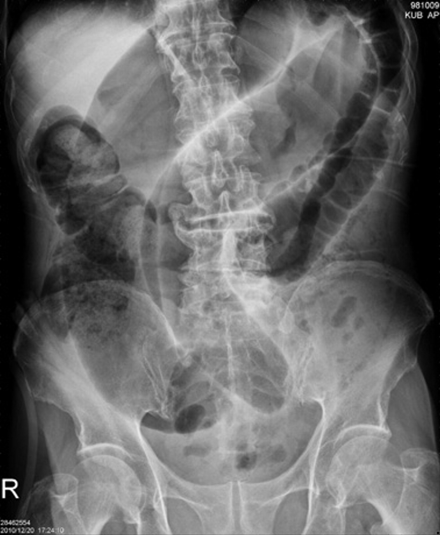
Sigmoid volvulus - 'coffee bean' sign. Here is another example of the 'coffee bean' sign of sigmoid volvulus. Sigmoid volvulus with coffee bean sign as seen on abdominal X-ray. Sigmoid volvulus is due to a twist at the base of the sigmoid mesentery which is a fixed position in the left iliac fossa.
View Coffee Bean Sign Images
The coffee bean sign is a radiological sign used to describe the twisting of the sigmoid colon about its mesenteric axis, mimicking the picture of a coffee bean (Figure 1C).The two side parts of the bean represent the gas‐filled segments of the dilated bowel creating an inverted U‐shape, whereas the central cleft of the bean represents the double thickness of opposed bowel walls. 1 The.

coffee bean sign meddic
The Coffee Bean Sign in Sigmoid Volvulus. Umer Salati et al., Radiology, 2011. Richard Louis Bean, MD. Paul A. Mori, Radiology, 2006. Findings of Cecal Volvulus at CT. Juliana M. Rosenblat et al., Radiology, 2010. A virus-derived microRNA-like small RNA serves as a serum biomarker to prioritize the COVID-19 patients at high risk of developing.

Whole Bean Coffee Sign Coffee signs, Coffee beans, Raw coffee beans
'COFFEE BEAN' SIGN AND 'WHIRL' SIGN. The preliminary view on abdominal computed tomography (CT) showed a distended sigmoid loop with an inverted U-shape, also known as the coffee bean sign, bent innertube sign, or kidney bean sign . This feature was also seen on plain abdominal radiography.

Coffee Bean Sign In Sigmoid Volvulus tech design trick
The coffee bean sign. An 89-year-old woman presented to our emergency department (ED) with a 4-day history of abdominal pain and vomiting. The physical exam revealed hypotension and a distended abdomen with generalized tenderness. Laboratory studies revealed hyperkalemia and elevated creatinine, lactate, and troponin.

Coffeebean sign CMAJ
"Coffee Bean," "Bird Beak," and "Whirl" Signs On axial CT images, cecal volvulus is suggested by the extreme dilatation of the cecum. When seen on conventional radiographs or tomograms, the cecal volvulus is seen as a rounded focal collection of air-distended bowel with haustral creases in the left upper quadrant that resembles a coffee bean (Figs. 2A , 2B , 2C and 3A , 3B ).

HD限定 Coffee Bean Sign Radiology さるあねか
coffee bean sign 5. Frimann-Dahl sign - three dense lines converge towards the site of obstruction. absent rectal gas 5. liver overlap sign. northern exposure sign. Fluoroscopy. Although now uncommonly performed, a water-soluble contrast enema exquisitely demonstrates this condition, with the appearances described as the beak sign (or bird beak.

The Coffee Bean Sign and Sigmoid Volvulus in an Elderly Adult Ladizinski 2013 Journal of
The presence of the coffee bean sign is pathognomonic of sigmoid volvulus. Coffee bean sign: Its meaning and importance Clin Case Rep. 2020 Jun 26;8(10):2086-2087. doi: 10.1002/ccr3.3064. eCollection 2020 Oct. Authors Eliza Stavride 1 , Charalampos Plakias 1 Affiliation 1.

Coffee Beans Yard Sign by WickedDesigns4
The "coffee bean sign" can be observed in imaging examinations. Although HD is most often diagnosed in the perinatal period and infancy, it can present during childhood, adulthood, and even in old age. SV may lead to difficult-to-control BP, which can increase short-term risk. Then, the management of SV is to relieve the obstruction and.
Coffee Bean Sign radRounds Radiology Network
A plain abdominal X-ray demonstrated a coffee bean sign indicating a sigmoid volvulus. A consequent CT scan of the abdomen revealed a deep outlet obstruction with massively dilated, elongated and twisted loop of the sigmoid colon and no signs of perforation. We performed emergency colonoscopy under the assumption of an acute sigmoid volvulus.
Coffee Bean Sign radRounds Radiology Network
An abdominal X-ray may show the classic coffee bean sign. Medical management includes stabilising the patient if they are shocked and correcting any electrolyte disturbances. A rigid sigmoidoscope can inspect the mucosa and place a flatus tube under direct vision, allowing gas and liquid stool to pass.

"Coffee Bean Sign" shown by abdominal CT scan. Download Scientific Diagram
Sigmoid volvulus is an important underlying cause of intestinal obstruction, which usually causes continuous and severe abdominal pain. Typical radiographic findings of this condition include the 'coffee bean sign', showing the presence of a U-shaped, distended sigmoid colon. 1 The treatment strategy includes endoscopic detorsion and surgery. 2 Minimal symptoms and the absence of.
Coffee beans signs logo template Royalty Free Vector Image
The coffee bean sign. Radiology 2000;216(1):178-179.. Sigmoid volvulus 'coffee-bean appearance' Dina HassanSheko. 2018 | Visual Journal of Emergency Medicine, Vol. 13. Acute Care Surgery Handbook. ChasenCroft, DougKwazneski, FrederickMoore. 2016. CLINICAL STUDY OF SIGMOID VOLVULUS.